
 ru
ru ge
ge ua
uaMarijuana as seen through the microscope
Bob Marley
Ford McCann is a modern-time enthusiastic botanist. Available online, his publication "Marijuana Under the Microscope", is a marvel of detailed macrophotography of cannabis herb under the microscope. Those pictures were taken with electronic micro-optics devices.
Marijuana seed trough a microscope
This image shows an enlarged Cambridge S200 electron microscope image of the surface of a hemp seed. The ridges on the seed are approximately 0.2 mm wide, they are used by the seed for absorbing moisture and also preventing bacterial infection.
Marijuana leaf under a microscope
The presented pictures are colored in Photoshop for easy viewing. In nature the colors of the captured elements are, of course, different shades of green.
Trichomes under a microscope
The oblong sharp shapes on the picture are nothing but trichomes growing on the end of the leaf. A fragment of a leaf measuring 0.2 mm in width is captured here.
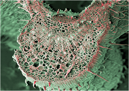
A leaf vein in section
This is the main vein of the leaf, pictured in section. Inside the vein are hemp pulp cells, which under the microscope form a complex three-dimensional structure. Their function is to transport nutrients. A plant fragment about 3 mm in diameter is captured.

The base of a leaf
The base of a leaf (petiole) in section. The notch in the image is an upward-facing vessel that scientists believe is responsible for transporting moisture in the leaf. The central part of the petiole contains the pulp cells. This is a panoramic image obtained from 18 separate images.

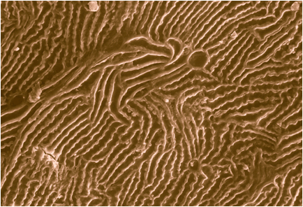
The underside of the leaf
A fragment about 3 mm wide is captured. In this photo of marijuana under a microscope, the round trichomes and needle-like trichomes that secrete THC molecules are clearly visible.

A carbon entry valve
The photo shows the closing cell of a marijuana plant under a microscope, in other words, the carbon entry valve.
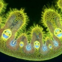
Cannabis bud under the microscope
The leaves of cannabis, as you may have already seen, are a complex multi-level structure. However, the main interest for fans of the plant is, of course, its flowers.
This photo of a cannabis inflorescence under a microscope shows different types of trichomes in different colors. Tetrahydrocannabinol can be up to 8% of the inflorescence mass.

Pistil of the female inflorescence

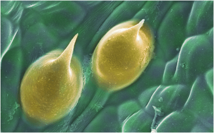
Glandular trichome
The glandular trichome is about 0.05 mm tall. Nevertheless, with these tiny hairs, visible only under an electron microscope, cannabis produces THC molecules.
And now a fun picture just to make you smile. This is how some people visualize cannabis under a microscope:

25.03.2017
Featured


